.jpg)
Anatomy
Origin:
The trigeminal motor nucleus is located in the mid pons tegmentum
and the trigeminal sensory nucleus extends the length of the brain stem and upper cervical cord. Its subnuclei include the principal sensory nucleus located in the pontine tegmentum, the mesencephalic nucleus extending into the midbrain and the spinal nucleus extending through the medulla and into the cord.
Fibers that convey touch and pressure terminate in the principal nucleus and fibers that convey pain and temperature terminate in the spinal nucleus.
Proprioceptive fibers from the muscles of mastication and the temporomandibular joint have their cell bodies in the mesencephalic nucleus.
Course:
It emerges from the anterior aspect of the pons as a small motor and a larger sensory root, the sensory root expands to form the flattened and crescent shaped trigeminal ganglion located lateral to the cavernous sinus.
Divisions:
There are three divisions of the trigeminal nerve arising from the trigeminal ganglion.
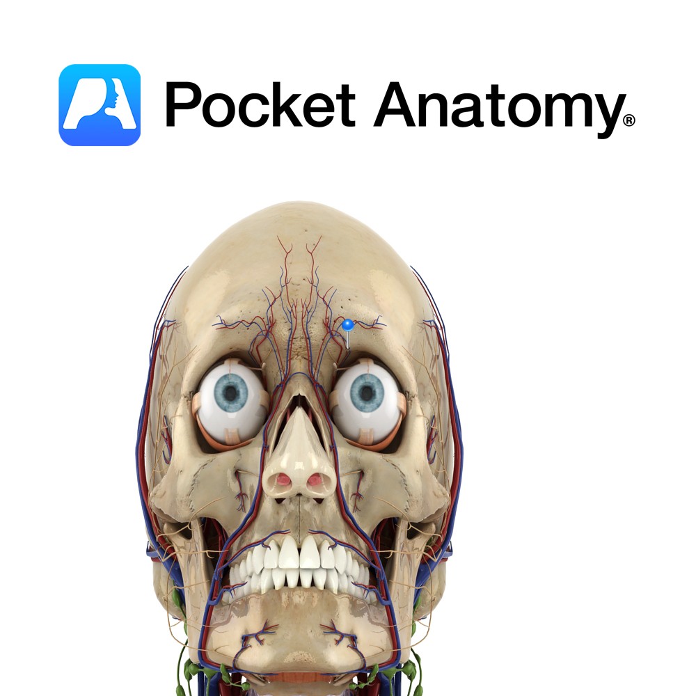
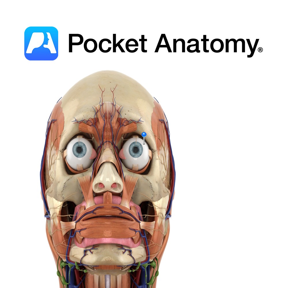
1) The ophthalmic nerve (CN V1) contains only sensory fibers and exits the skull via the superior orbital fissure to enter the orbital cavity.
2) The maxillary nerve (CN V2) contains only sensory fibers. It exits the skull through the foramen rotundum. The pterygopalatine ganglion is associated with the maxillary nerve and innervates the lacrimal gland and nasal and oral mucous membranes.
3) The mandibular nerve (CN V3) contains both sensory and motor fibers. It exits the skull via the foramen ovale.
Functions
The ophthalmic nerve (CN V1) supplies sensation from the skin, mucous membranes and conjunctiva of the front of the head and nose.
The maxillary nerve (CN V2) supplies sensation from the skin over the maxilla, upper lip, teeth and palate and the mucous membranes associated with the upper jaw.
The mandibular nerve is motor to the muscles of mastication, the mylohyoid, anterior belly of the digastric muscle, tensor veli palatini and tensor tympani muscle. It provides sensation from over the mandible, lower teeth, temporomandibular joint, mucosa of the mouth and the anterior two thirds of the tongue.
Clinical
Injury to CN V can cause:
– Paralysis of the muscles of mastication.
– Loss of soft tactile, thermal, or painful sensations to the face.
– Loss of the corneal reflex and the sneezing reflex.
The corneal reflex, also known as the ‘blink reflex’, is when the eye blinks in response to the touching of the cornea. The afferent fibers of the reflex arc are supplied by the ophthalmic nerve (CN V1) which receives impulses from the cornea, and the efferent limb of the reflex arc is supplied by CN VII to the orbicularis oculi muscles.
Injury to CN V may occur to any or all of its branches during its course.
The spinal nucleus of V is typically involved in the lateral medullary syndrome of Wallenberg (resulting in vertigo, nausea, vomiting, ipsilateral cerebellar signs, ipsilateral Horner syndrome, ipsilateral palatal, pharyngeal, and vocal cord paralysis (due to involvement of the nucleus ambiguus), ipsilateral facial hypalgesia and thermoanaesthesia, contralteral trunk and limb hypalgesia and thermoanaesthesia, and occasioanlly, hiccups).
CN V1 may also be involved in cavernous sinus syndromes (which will be associated with any or all of ipsilateral palsies of CN III, IV, and VI, and Horner’s syndrome).
Injury to the trigeminal nerve has many potential etiologies including trauma, tumours, aneurysms and meningeal infections.
Interested in taking our award-winning Pocket Anatomy app for a test drive?
![]()