Functions
Allows communication between oral and nasal cavity. Separates (with epiglottis) food/drink and air into oesophagus and trachea respectively.
Helps (along with nostils and nasal cavity) to warm and humidify inhaled air before lungs.
Important area for reverberation when vocalising.
Anatomy
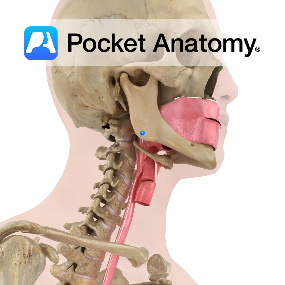
Cone shaped area at back of throat, typically 3.6cm2 cross section in men, 3.2cm2 in women (varies when breathing by 0.6cm2 and 0.1cm2, respectively). Situated posterior to nasal and oral cavities, superior to oesophagus and larynx. Divided into nasal, oral, or laryngeal pharynx with divisions of oropharyngeal isthmus and epiglottis.
Consists of outer circular muscles (inferior, middle and superior constrictor) which propel food bolus down during swallowing, and inner longitudinal muscles (stylopharyngeus, salpingopharyngeus, palatopharyngeus) which shorten and widen pharynx when swallowing.
Supplied by superior thyroid, lingual, and ascending pharyngeal arteries. Innervated by vagus and glossopharyngeal nerves.
Contains: Pharyngeal tonsils (nasal part), Waldeyer’s Ring (nasal and oral part), Palatine tonsils (oral part), eustachian tubes.
Clinical
HACEK organisms (Haemophilus species, Aggregatibacter, Cardiobacterium, Eikenella, Kingella) are a group of fastidious Gram-negative bacteria that naturally colonise the oral-pharynx. They are a common cause of infective endocarditis, especially in young children. The treatment of choice for these organisms is ceftriaxone, a third generation cephalosporin.
Interested in taking our award-winning Pocket Anatomy app for a test drive?
![]()