Anatomy
Origin:
Anterior and lateral surfaces of the proximal two-thirds of the femoral shaft.
Insertion:
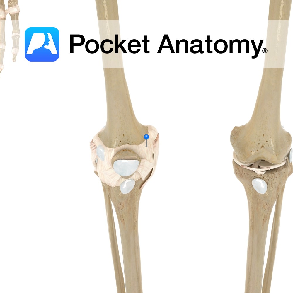
Lateral margin and base of the patella via the quadriceps femoris tendon. The quadriceps femoris tendon is functionally continuous with the patellar ligament which runs from the apex of the patella to the tibial tuberosity.
Key Relations:
One of the five muscles of the anterior compartment of the thigh.
Note:
The quadriceps femoris is a large muscle group containing the 3 vastus muscles and the rectus femoris muscle of the anterior thigh. The quadriceps femoris tendon represents their shared insertion onto the patella.
Functions
Together with the other muscles that make up quadriceps femoris extends the leg at the knee joint.
Supply
Nerve Supply:
Femoral nerve (L2, L3, L4).
Blood Supply:
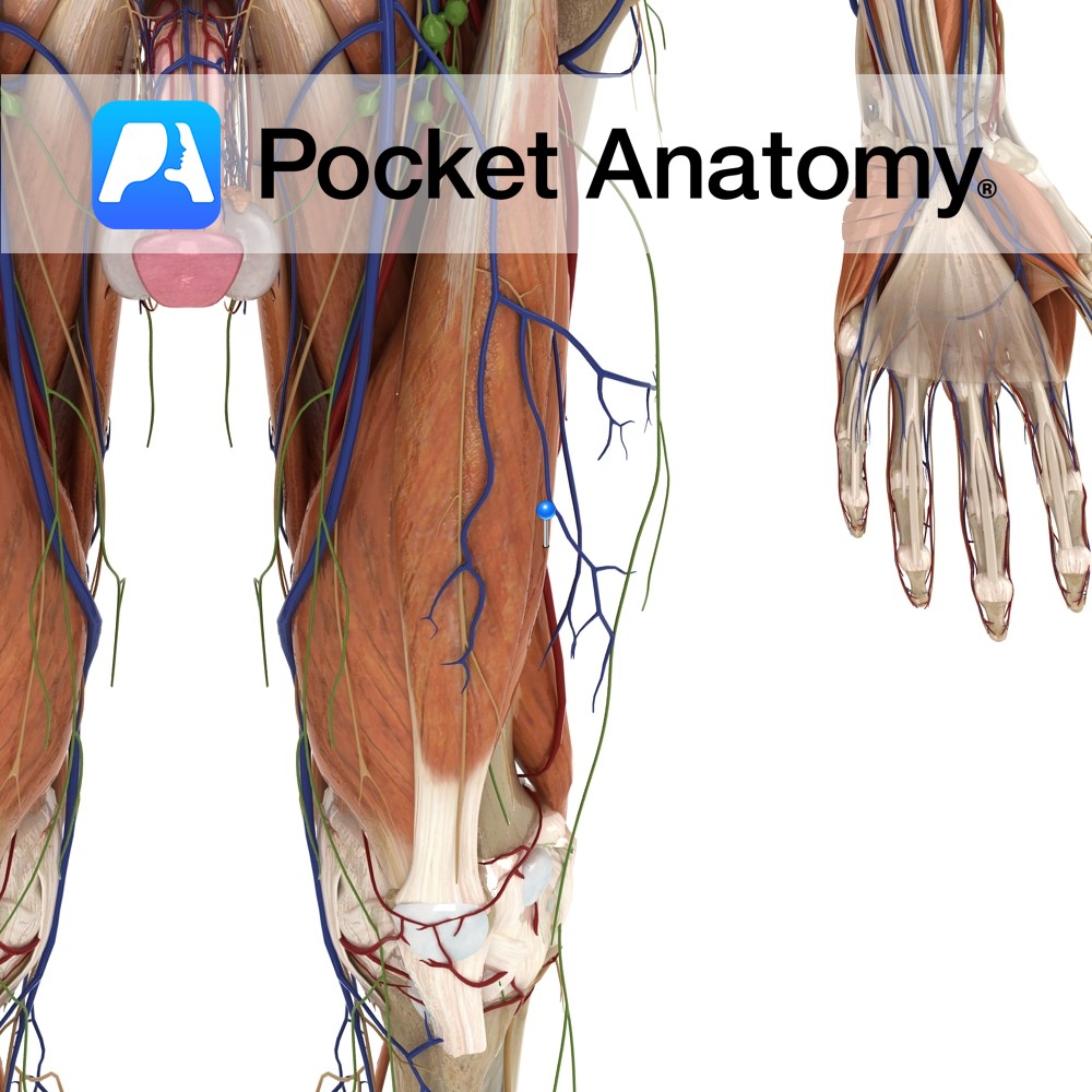
-Lateral circumflex femoral artery
-Deep femoral artery.
Clinical
Traction injury of the insertion of the quadriceps tendon referred to as Osgood-Schlatter disease is a common condition in adolescents after strenuous activity. Repetitive contraction of the quadriceps muscle can cause strain to the patellar ligament and cause damage and tearing at its attachment to the tibial tuberosity. As callous is layed down during healing the tibial tubercle becomes pronounced. Bony prominence of the tibial tubercle would be a characteristic finding upon examination, as well as a patient complaining of anterior knee pain getting worse over time.
Interested in taking our award-winning Pocket Anatomy app for a test drive?
![]()