PocketAnatomy® is a registered brand name owned by © eMedia Interactive Ltd, 2009-2022.
iPhone, iPad, iPad Pro and Mac are trademarks of Apple Inc., registered in the U.S. and other countries. App Store is a service mark of Apple Inc.
Anatomy Origin: Superficial head (tendinosus): Lateral epicondyle of humerus, Radial collateral and annular ligaments Deep head (muscular): Supinator crest and fossa of ulna. Insertion: Lateral aspect of the proximal third of the radius. Key Relations: -The posterior interosseus nerve travels between the two heads of supinator as it enters the posterior forearm. -One of the
- Published in Pocket Anatomy Pins
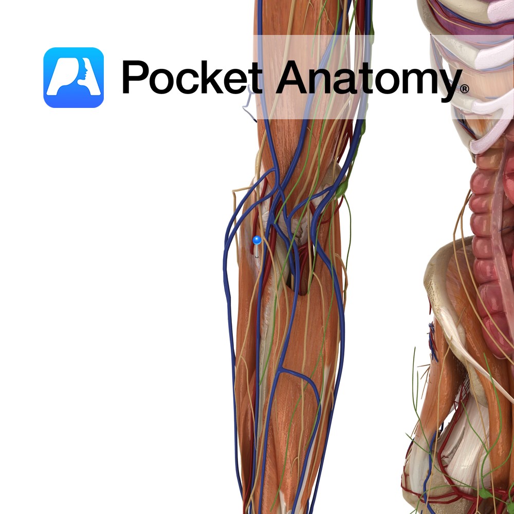
Anatomy Course Branches from the brachial artery about a third of the way along the humerus. It travels along the medial head of the triceps towards the elbow joint, where it anastomoses with the posterior ulnar recurrent artery. Supply Contributes to the blood supply of the medial aspect of the arm and to the elbow
- Published in Pocket Anatomy Pins
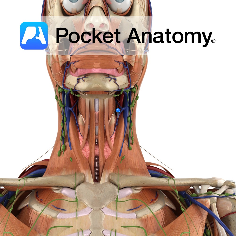
Anatomy Course Begins in the superior region of the thyroid. It emerges from the thyroid to run laterally, crossing the common carotid artery and draining into the internal jugular vein. Drain Drains the superior region of the thyroid gland. Interested in taking our award-winning Pocket Anatomy app for a test drive?
- Published in Pocket Anatomy Pins
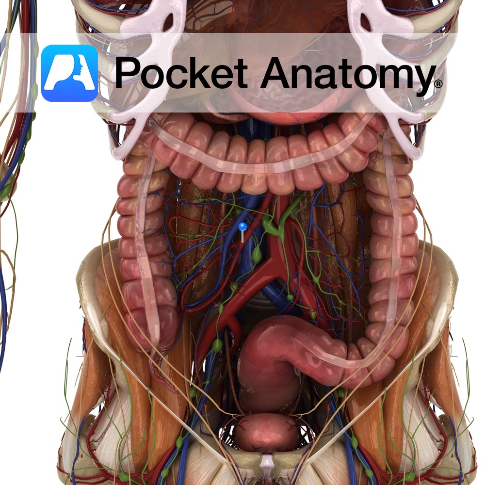
Anatomy Course Begins as tributaries from the venous plexus around the rectum and the sigmoid colon. It ascends in the pelvis and crosses the internal iliac vein to become the inferior mesenteric vein. Drain Drains the rectum and sigmoid colon. Interested in taking our award-winning Pocket Anatomy app for a test drive?
- Published in Pocket Anatomy Pins
Anatomy Course One of the terminal branches of the inferior mesenteric artery. It descends in the sigmoid mesocolon and crosses over the left internal iliac artery. It divides at the vertebral level of SIII, and continues to descend by the rectum where it anastomoses with the middle and inferior rectal arteries. Supply Supplies the rectum.
- Published in Pocket Anatomy Pins
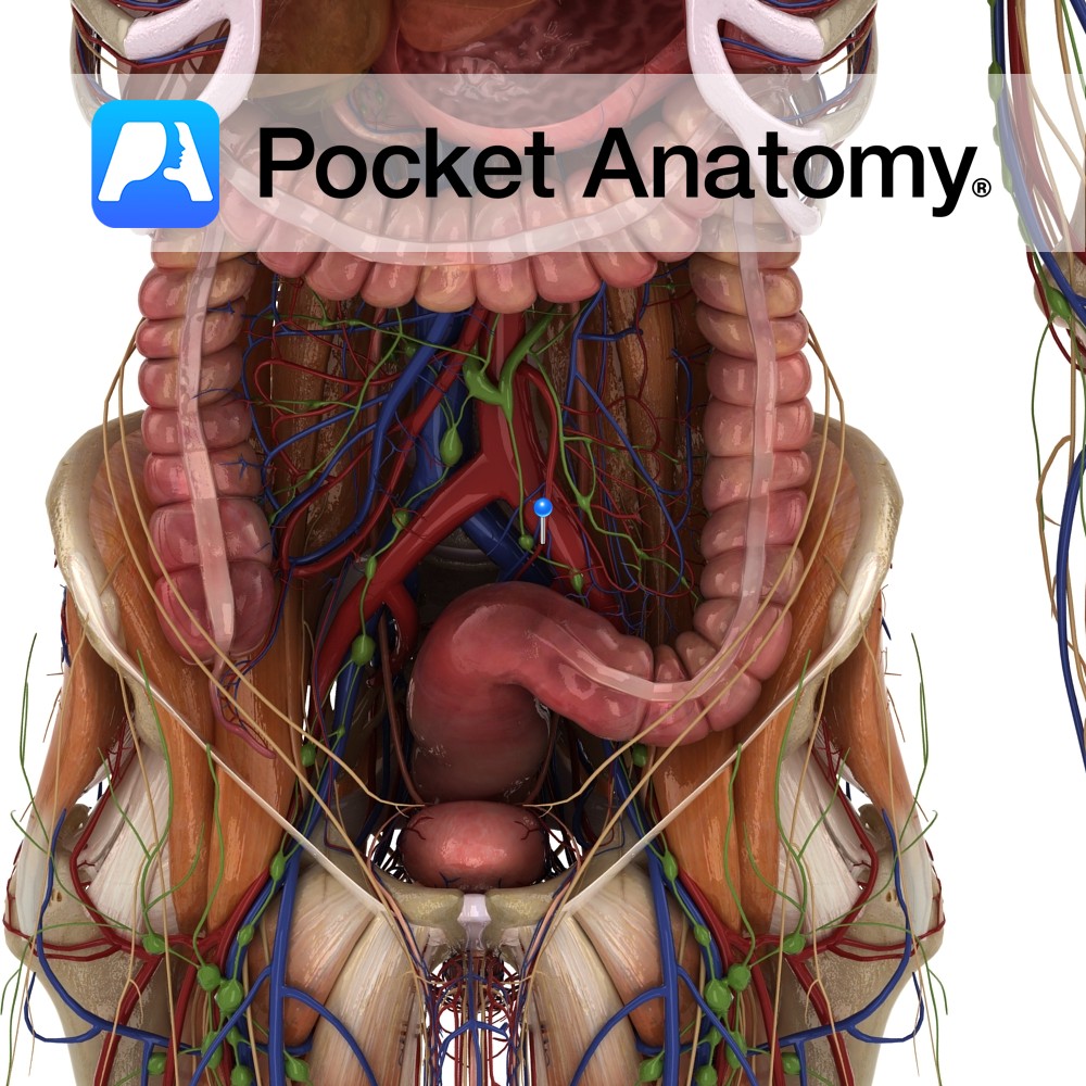
Anatomy Course Formed by the union of several veins in the abdomen that drain the region known as the embryonic midgut. It is formed in the mesentery, through which it ascends and then travels up behind the pancreas. Here it joins with the splenic vein to form the portal vein. Drain Drains the region of
- Published in Pocket Anatomy Pins
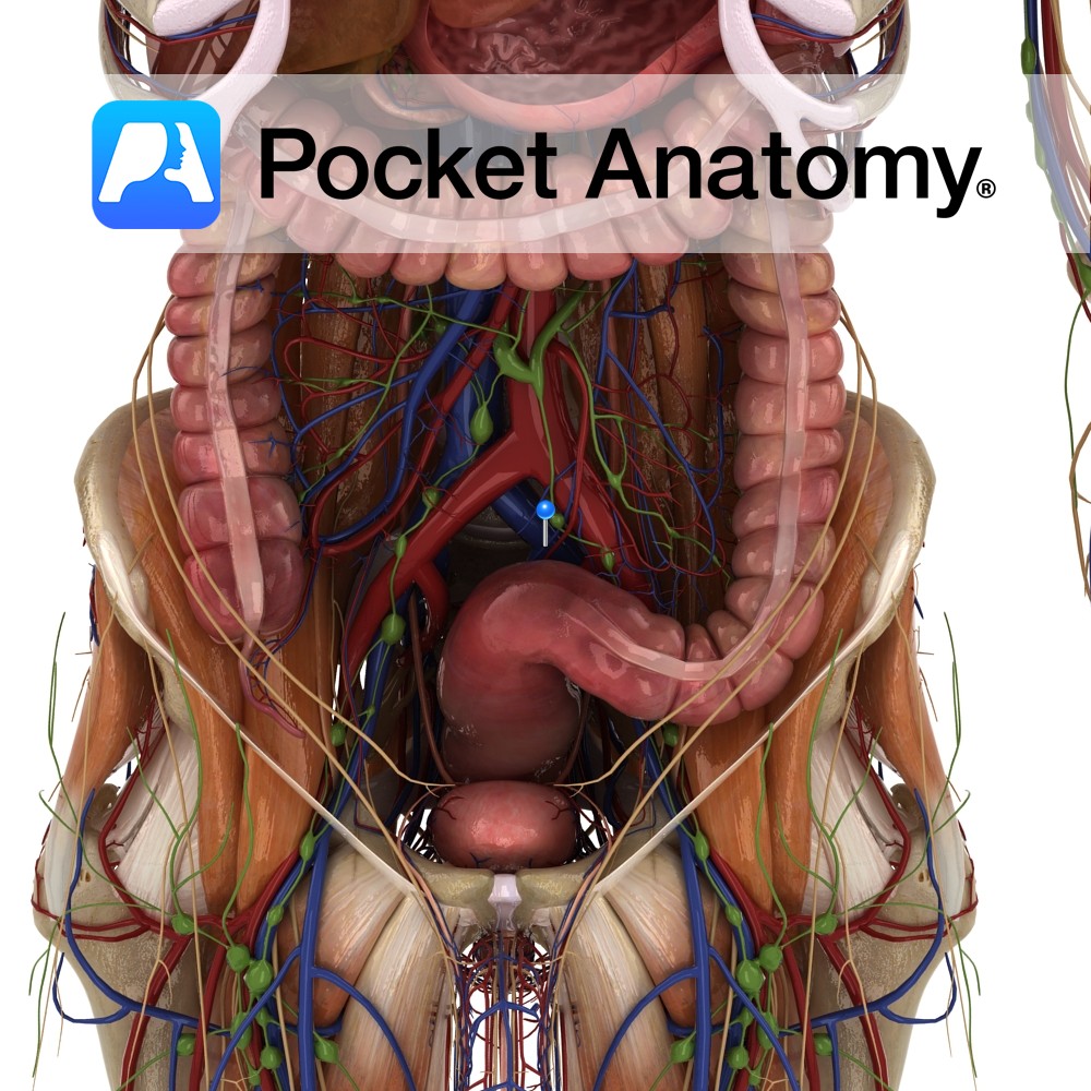
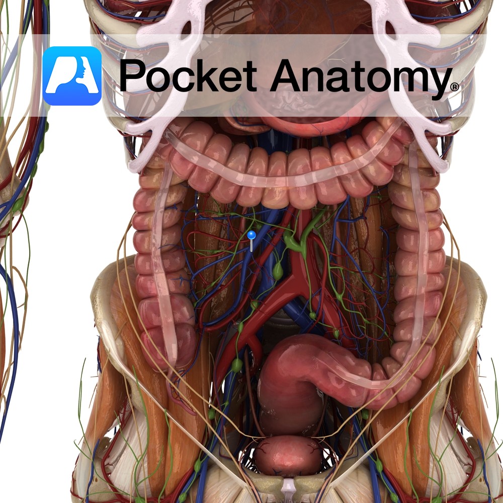
Anatomy Course An anterior branch of the abdominal aorta that arises at approximately the vertebral level of L1. It descends into the abdomen, passing in front of the left renal vein and the inferior duodenum and passing behind the neck of the pancreas and the splenic vein. It also crosses in front of the uncinate
- Published in Pocket Anatomy Pins
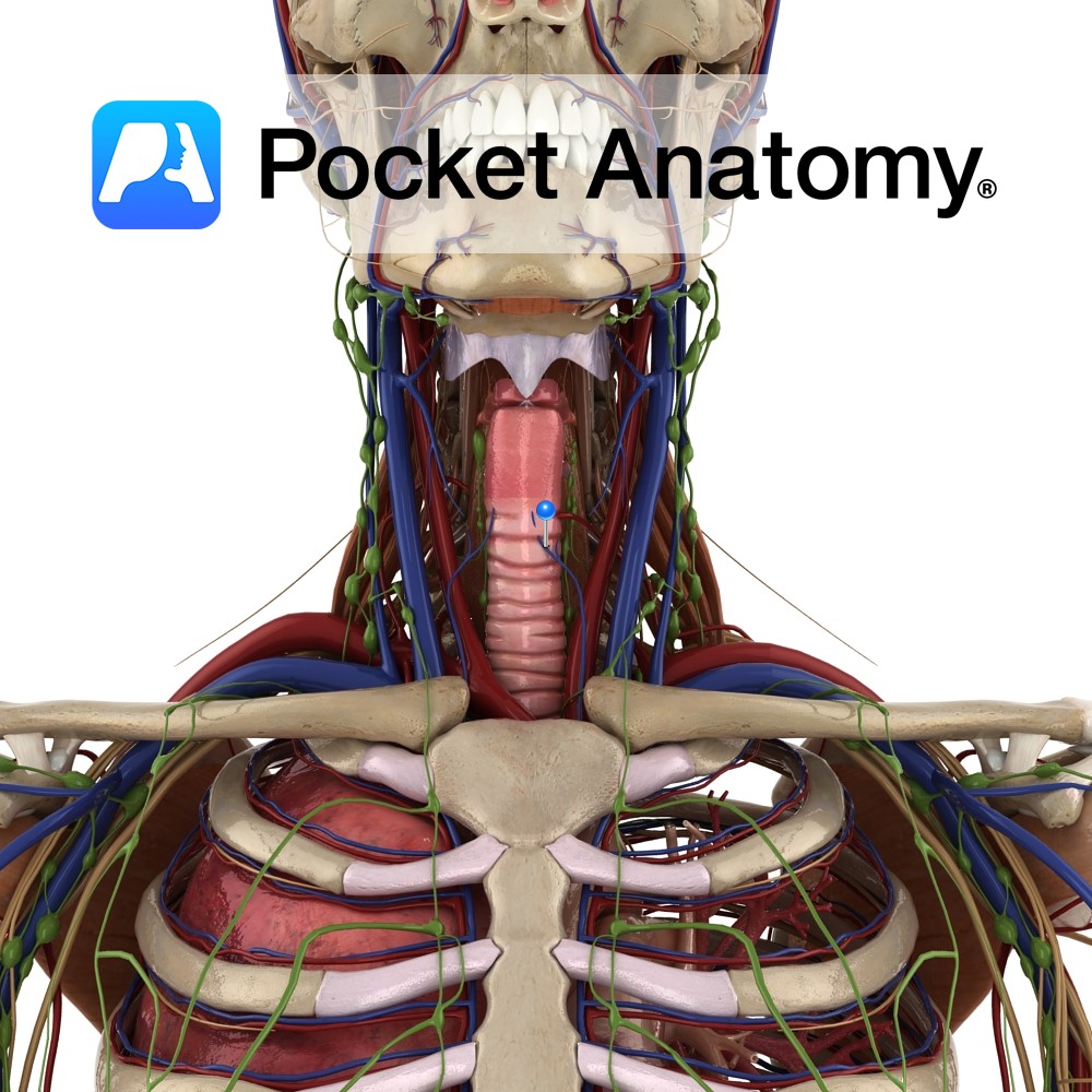
Anatomy Course Begins from a network of veins within the larynx. It exits the larynx by piercing the hyothyroid membrane and drains into the superior thyroid vein. Drain Drains the larynx and its mucous membranes. Interested in taking our award-winning Pocket Anatomy app for a test drive?
- Published in Pocket Anatomy Pins
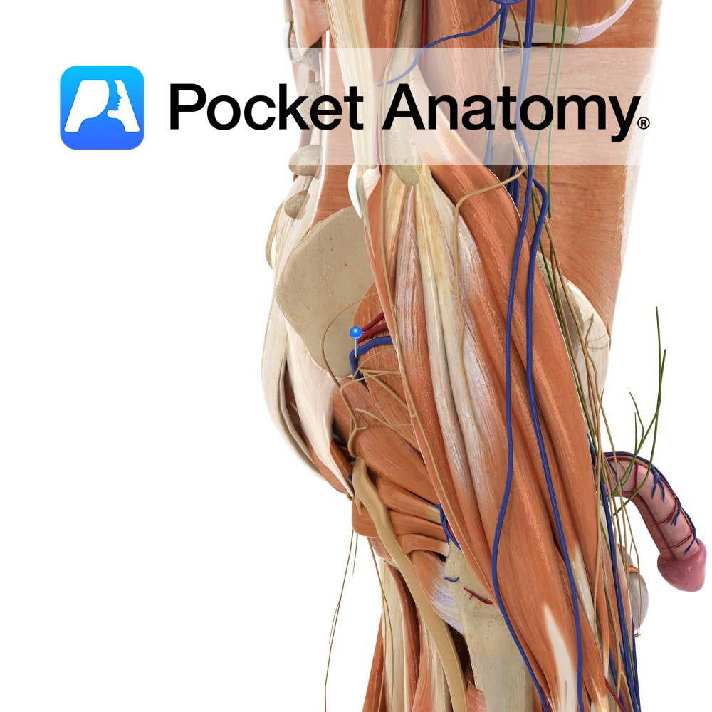
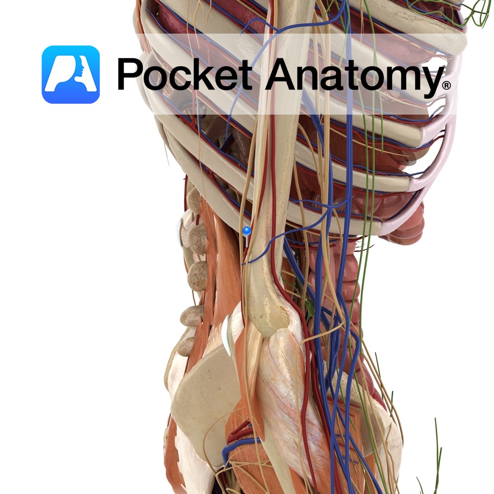
Anatomy Course Begins in the gluteal region, where a number of smaller veins come together. It travels towards the greater sciatic foramen, where it enters the pelvic cavity. It travels along the posterior pelvic wall to drain into the internal iliac vein. Drain Drains the gluteal region. Interested in taking our award-winning Pocket Anatomy app
- Published in Pocket Anatomy Pins
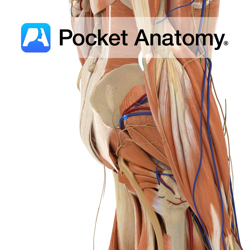
Anatomy Course A terminal nerve of the lumbosacral plexus. It is formed by fibres from spinal segments L4 – S1. It exits the pelvic cavity by passing through the greater sciatic foramen, above the piriformis muscle. It travels anterolaterally between gluteus medius and minimus, and terminates in the tensor fasciae latae muscle. Supply The superior
- Published in Pocket Anatomy Pins