PocketAnatomy® is a registered brand name owned by © eMedia Interactive Ltd, 2009-2022.
iPhone, iPad, iPad Pro and Mac are trademarks of Apple Inc., registered in the U.S. and other countries. App Store is a service mark of Apple Inc.
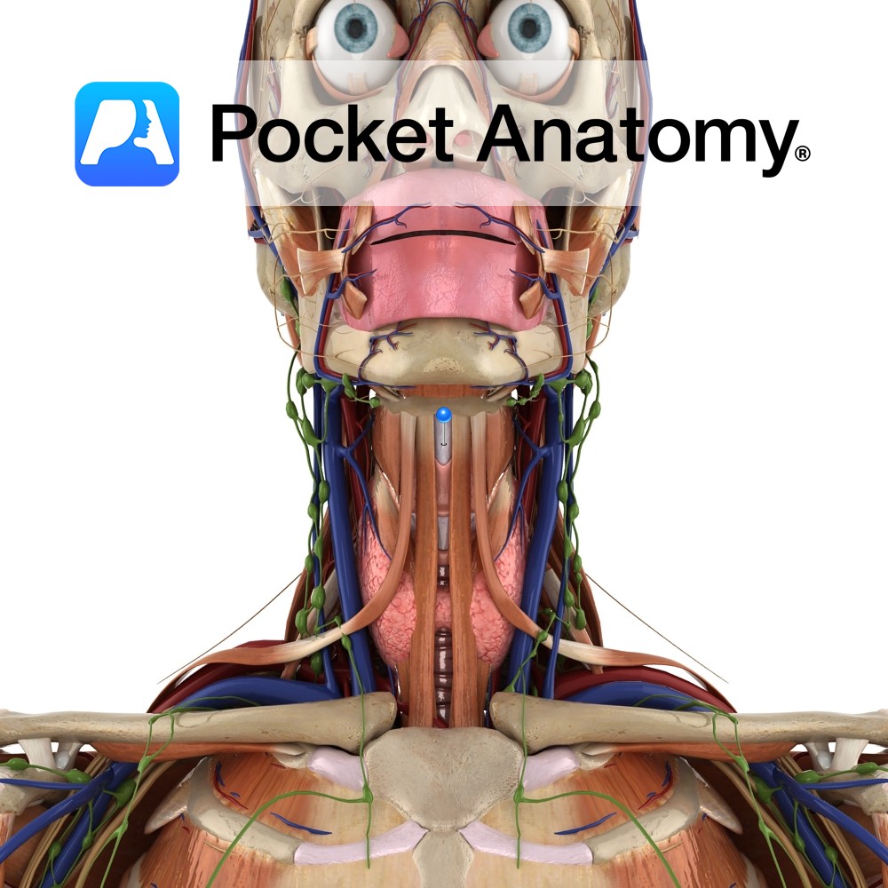
Anatomy Connects the superior borders of the thyroid laminae and superior horns to the hyoid bone above. It is thickened in the midline as the median thyrohyoid ligament. Functions Helps to hold the thyroid in place. Interested in taking our award-winning Pocket Anatomy app for a test drive?
- Published in Pocket Anatomy Pins
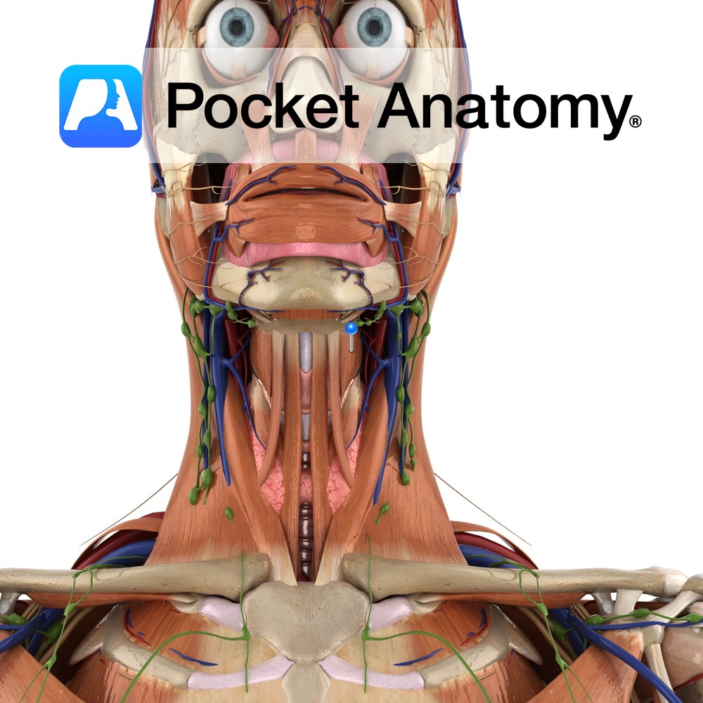
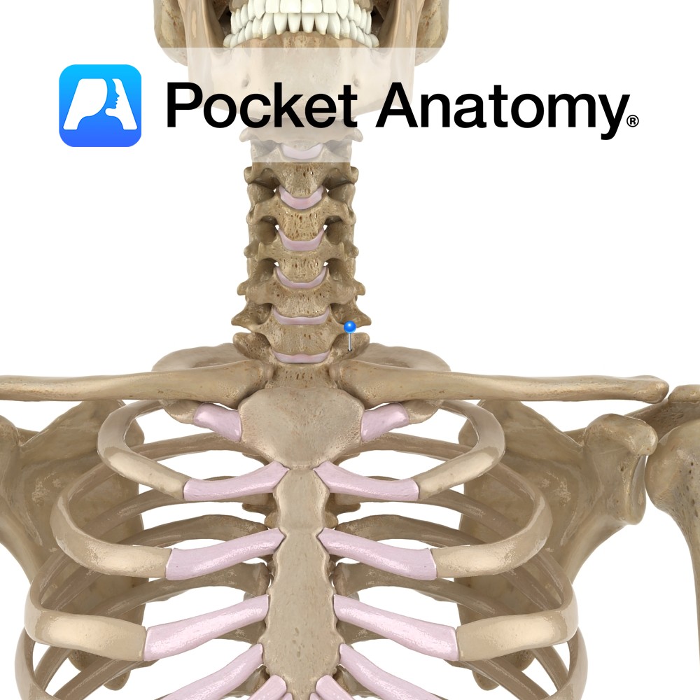
Anatomy Origin: Oblique line on lamina of thyroid cartilage. Insertion: Body of hyoid bone. Key Relations: -Is one of the infrahyoid muscles (sometimes referred to as ‘strap muscles’) lying in the muscular triangle of the neck. -Lies deep to omohyoid and sternohyoid. Functions -Depresses the hyoid bone. -When hyoid is fixed, raises the larynx e.g.
- Published in Pocket Anatomy Pins
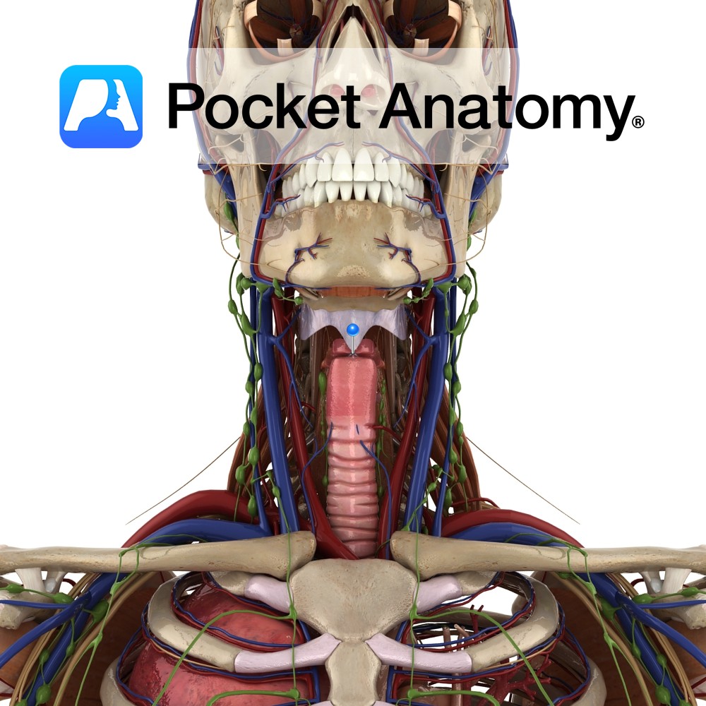
Anatomy Attaches from the inferior end of the epiglottis to the thyroid just below the thyroid notch. Functions Attaches the epiglottis to the thyroid. It also helps hold it in place. Interested in taking our award-winning Pocket Anatomy app for a test drive?
- Published in Pocket Anatomy Pins
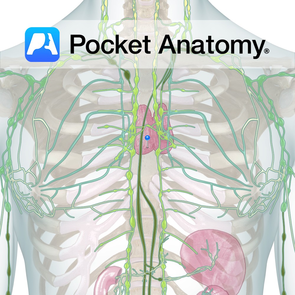
Anatomy Finishing school, turns Lymphocytes from Bone Marrow into immunocompetent T(hymus)-Lymphocytes and sends them out into the Lymphatic world (to various Lymph Nodes/Glands), enlarges from 12/40 fetus to puberty then shrinks. Situated behind upper part Breastbone (Manubrium Sterni), bi-lobed with junction at front trachea, lobes fat- and WBC-rich. Clinical Fundamental functions of Lymphatic System; tissue
- Published in Pocket Anatomy Pins
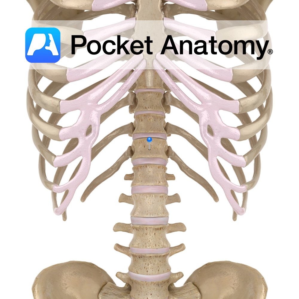
Anatomy Vertebrae T9 – T12 (lowermost thoracic) show progressive change from the mid-thoracic pattern of demi-facets on the body (whereby the heads of ribs straddle 2 vertebrae), to single facets on the pedicle. Also, the transverse process has no articular surface (and 12th rib has no tubercle). Clinical Articulates above with T11 (top of vertebral
- Published in Pocket Anatomy Pins
Anatomy Has a heart-shaped body, lateral surface (each side) of which has an articulating facet for the head of 1st rib and a demi-facet for the upper half of the head of 2nd rib. From either side of the back of the body, pedicles project posteriorly, joining laminae which themselves meet at the midline (from
- Published in Pocket Anatomy Pins
.jpg)
Anatomy The Thoracic Duct conveys lymph from 3/4 the body (all except the Right side of the head and neck, R upper limb and R side of chest), emptying into either the L Subclavian or L Brachiocephalic Vein. Extends from level L2 (where R and L Lumbar Trunks and Intestinal Trunk join to form the
- Published in Pocket Anatomy Pins
.jpg)
Anatomy Course The ascending portion of the thoracic aorta is that directly from the heart to the arch of the aorta. It begins at the base of the left ventricle of the heart, and is contained in the pericardium for its course. Supply All blood that is circulated throughout the systemic circulation passes through the
- Published in Pocket Anatomy Pins
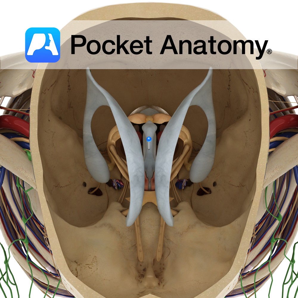
Anatomy Is a slit-like cavity lined with ependymal cells, located between the two thalami and is a part of the series of fluid-filled cavities, which make up the ventricular system. It is connected anteriorly to the lateral ventricles via the interventricular foramen of Monro. The anterior wall is formed by the lamina terminalis. The lateral
- Published in Pocket Anatomy Pins
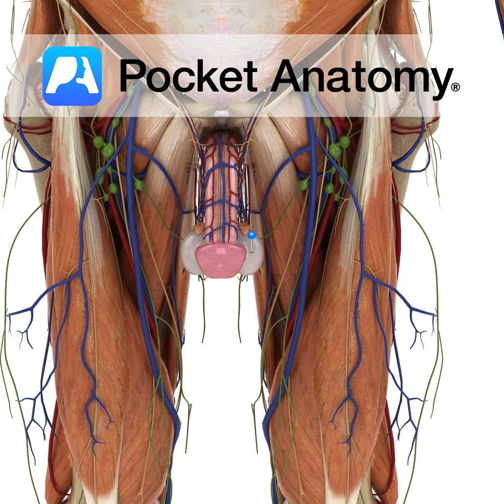
Anatomy Male gonad [organ that makes gametes – sperm – through a process (spermatogenesis) that involves a type of cell division (meiosis) that produces cells that are haploid, ie have half the chromosome complement of other – diploid – cells; female gamete is ovary, its meiotic product are eggs], produces sperm and testosterone, ellipsoid (about
- Published in Pocket Anatomy Pins