PocketAnatomy® is a registered brand name owned by © eMedia Interactive Ltd, 2009-2022.
iPhone, iPad, iPad Pro and Mac are trademarks of Apple Inc., registered in the U.S. and other countries. App Store is a service mark of Apple Inc.
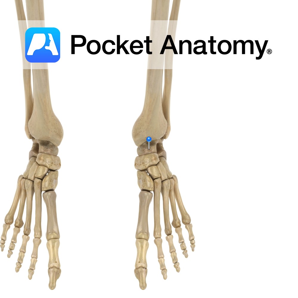
Anatomy Anklebone. Tarsal bone, irregular shape (boxing glove from above), transmitting weight of body to foot through articulations up with malleoli of tibia and fibula (talocrural joint – hinge), down with calcaneus (subtalar joint) and forward with navicular (along with calcaneocuboid joint, forms transverse talar joint). Clinical Fractures can be difficult to treat, can lead
- Published in Pocket Anatomy Pins
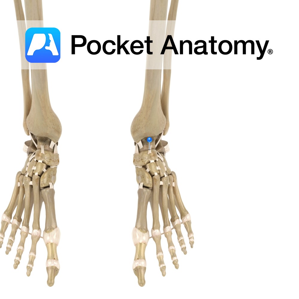
Anatomy A broad, thin ligament that connects the neck of the talus to the dorsal surface of the navicular. Functions Provides static stability to the talocalcaneonavicular joint. Interested in taking our award-winning Pocket Anatomy app for a test drive?
- Published in Pocket Anatomy Pins
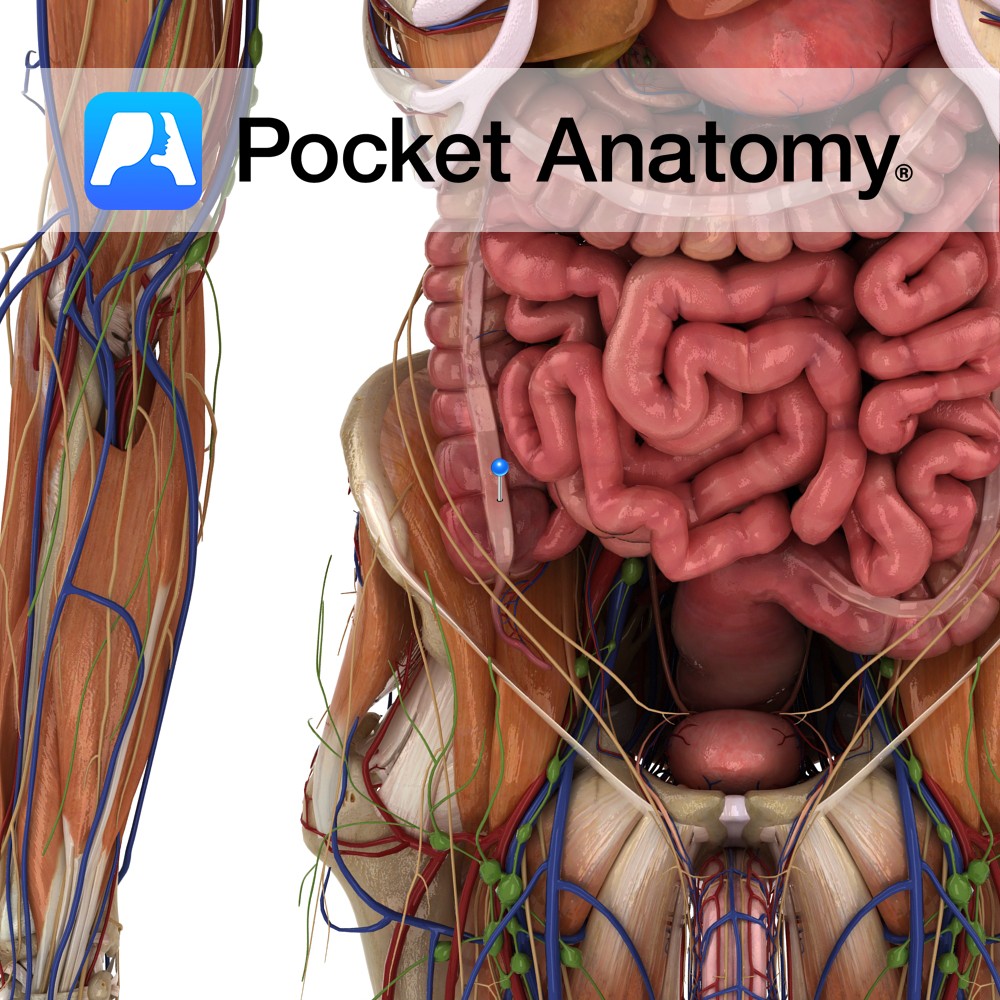
Anatomy 3 distinct, separated, equally spaced, longitudinal ribbons/straps of muscle (mesocolic, free, omental) along the length of ascending, transverse, descending and sigmoid colon, visible just below serosa, shorter than other corresponding colonic layers, thus puckering the colon between them (even more on contraction), and giving rise to haustra/sacculations; converge at either end – rectum, base
- Published in Pocket Anatomy Pins
-bone.jpg)
Anatomy Also known as extra-sutural bone. An extra, occasionally present, irregular bone, in a cranial suture. Most commonly seen in the lambdoid area. Vignette Ole Worm, 16th/17th century Danish physician. Also known as Inca bone; relatively high prevalence in Peruvian mummies. Presence of multiple wormians a marker in diagnosis osteogenesis imperfecta (Brittle Bone Disease). Interested
- Published in Pocket Anatomy Pins
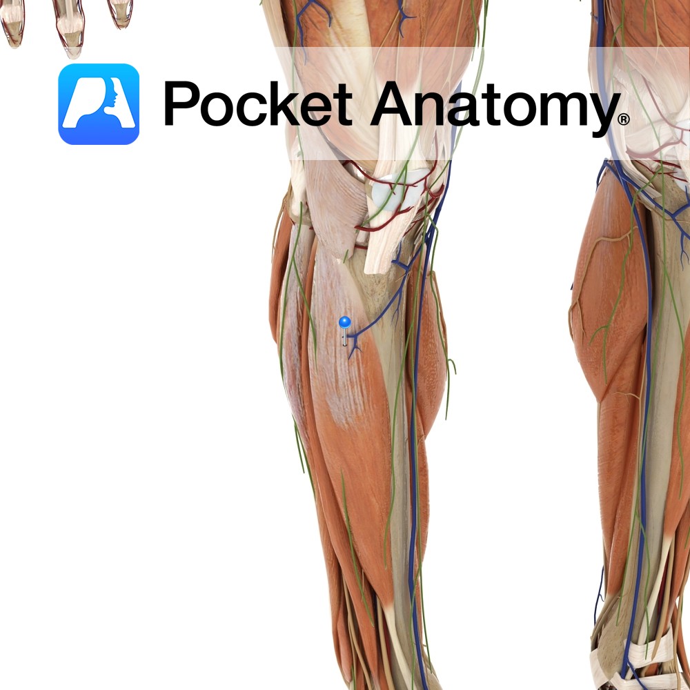
Anatomy Course Originates from the tibial nerve just above the knee joint, between the heads of the gastrocnemius muscle. It descends superficially to the gastrocnemius muscle, and penetrates the deep fascia about halfway down the calf, to run subcutaneously. It continues down the leg, and passes around the lateral malleolus to continue to supply the
- Published in Pocket Anatomy Pins
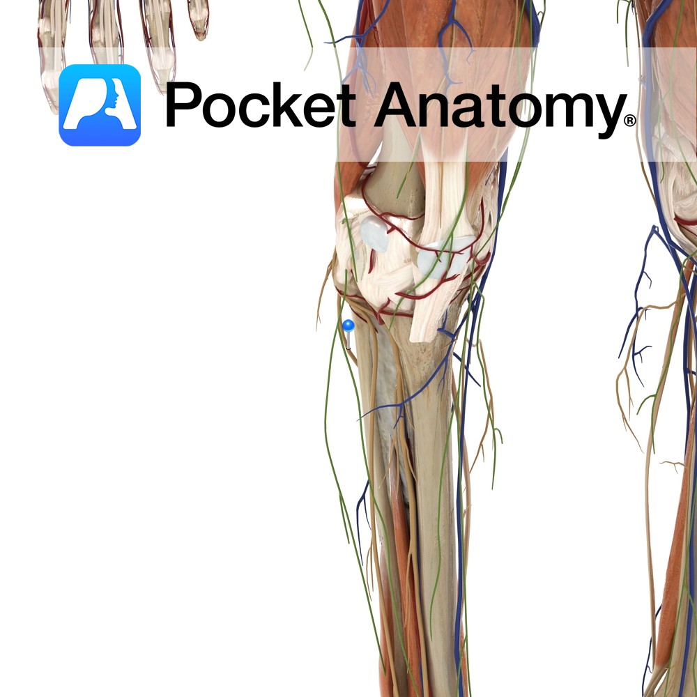
Anatomy Course The sural communicating branch of the common peroneal nerve contributes to the sural nerve. It arises from the common peroneal nerve in the popliteal space and passes over the lateral head of the gastrocnemius muscle. About halfway down the leg, it joins with the medial sural nerve to form the sural nerve. Supply
- Published in Pocket Anatomy Pins

Anatomy Course A terminal branch of the ophthalmic vein. Drain Drains forehead and front of scalp. Interested in taking our award-winning Pocket Anatomy app for a test drive?
- Published in Pocket Anatomy Pins

Anatomy Course A terminal branch of the ophthalmic artery (which is a branch of the internal carotid) – branches off where the ophthalmic artery travels posterior to the trochlea, exits the orbit medially, and ascends the forehead. Supply Along with the ophthalmic artery, it supplies the structures of the orbit of the eye, including the
- Published in Pocket Anatomy Pins
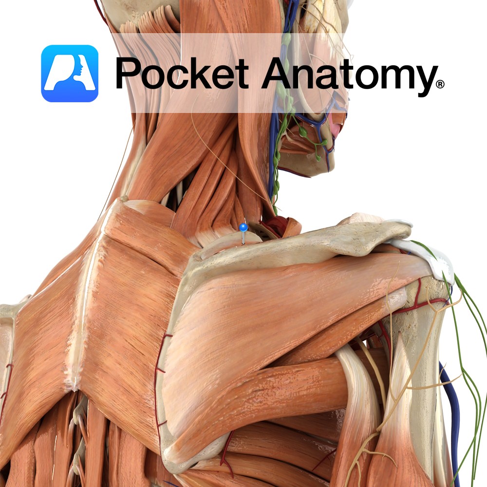
Anatomy Origin: Medial two-thirds of the supraspinous fossa of scapula. Insertion: Superior facet of greater tubercle of humerus. Key Relations: -The muscle forms a tendon that passes under the acromion and above the shoulder joint. -One of the four muscles of the rotator cuff muscle group. Functions -Initiates abduction of the arm to 15° at
- Published in Pocket Anatomy Pins
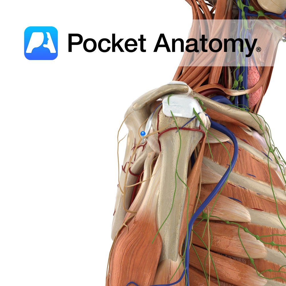
Anatomy Course Arises in the upper trunk of brachial plexus (formed by the union of C5 and C6). Passes downward, laterally (deep to the omohyoid and trapezius, then posteriorly beneath trapezius) before reaching the suprascapular notch. Travels beneath the suprascapular notch before dividing into two branches; one supplies supraspinatus, the other supplies infraspinatus. Supply Sends
- Published in Pocket Anatomy Pins