Anatomy
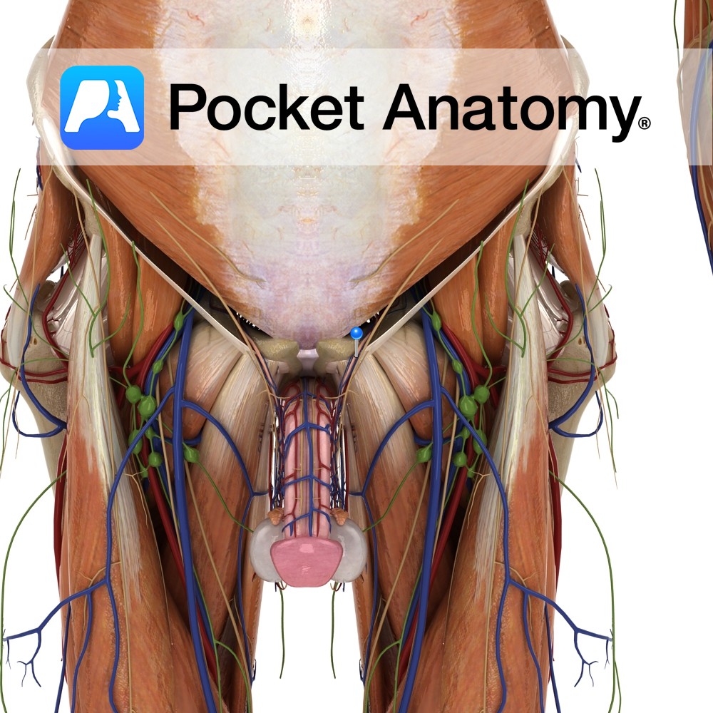
In development, the testes bulge/bubble down and out of the abdomen (to escape to a lower temperature), through a passage in the abdominal wall (the inguinal canal) bringing with them layers and structures (abnormal bulging is called herniation).
The Spermatic Cord reflects this abdominal origin and connects the testis to the abdomen; its fascias are extensions of those of the external/internal oblique muscles and of the transversalis fascia, its muscle (cremaster) originates in internal oblique muscle. Constituents of cord; “3, 3, 3, 3”. Arteries (3 – testicular, deferens, cremaster). Nerves (3 – genital, autonomic/visceral afferents, ilio-inguinal). Fascia (3 – external/internal spermatic, cremasteric). 3 other structures; pampiniform plexus, lymphatics, vas (ductus) deferens.
Physiology
Sperm travel up the cord, into the abdomen, from one part of the external genitalia (testes), re-emerging in and passing through and out of another (penis).
Clinical
The testis can twist on its cord (torsion), cutting off blood supply, producing severe pain of ischemia; ultrasound helps diagnosis. May resolve spontaneously or require emergency surgical release (within 6 hours).
Interested in taking our award-winning Pocket Anatomy app for a test drive?
![]()
.jpg)
.jpg)