
PocketAnatomy® is a registered brand name owned by © eMedia Interactive Ltd, 2009-2027.
iPhone, iPad, iPad Pro and Mac are trademarks of Apple Inc., registered in the U.S. and other countries. App Store is a service mark of Apple Inc.
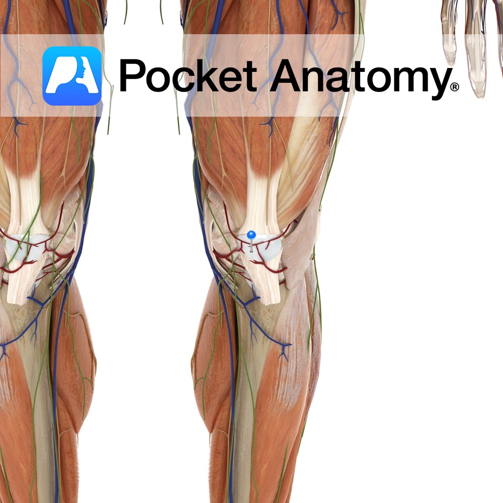
Anatomy Situated in front of the patella bone. Functions Acts as a cushion between bone and skin reducing friction between both. Clinical Prepatellar bursitis or ‘housemaid’s knee’ is a common injury to the bursa. The bursa becomes swollen due to repetitive work in a kneeling position. Interested in taking our award-winning Pocket Anatomy app for
- Published in Pocket Anatomy Pins
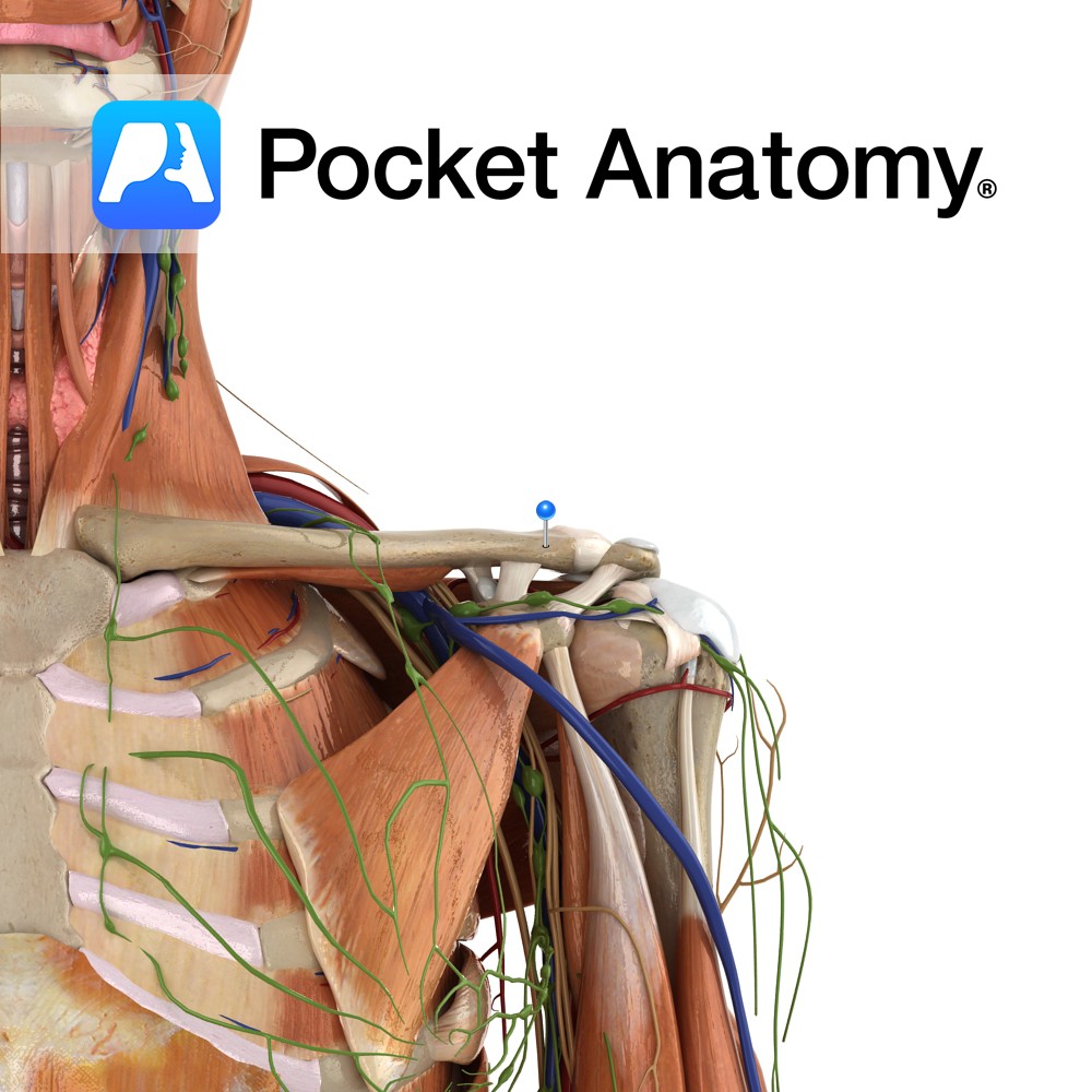
Motion The acromioclavicular joint is classified as a multiaxial synovial plane joint but it is more helpful to think of it as a pivot point. As the name suggests it is the articulation of the acromion of the scapula with the clavicle. It allows you to raise your arms above your head i.e. scapular elevation
- Published in Pocket Anatomy Pins
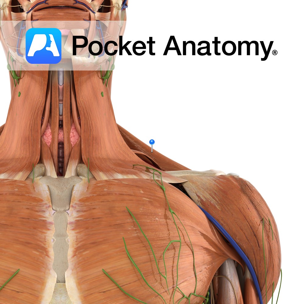
Anatomy Origin: Superior nuchal line of the occipital bone, external occipital protuberance, ligamentum nuchae and spinous processes of C7 to T12. Insertion: Superior fibres: posterior border of the lateral third of the clavicle. Middle fibres: medial border of the acromion and the superior edge of the spine of the scapula. Inferior fibres: tubercle of the
- Published in Pocket Anatomy Pins
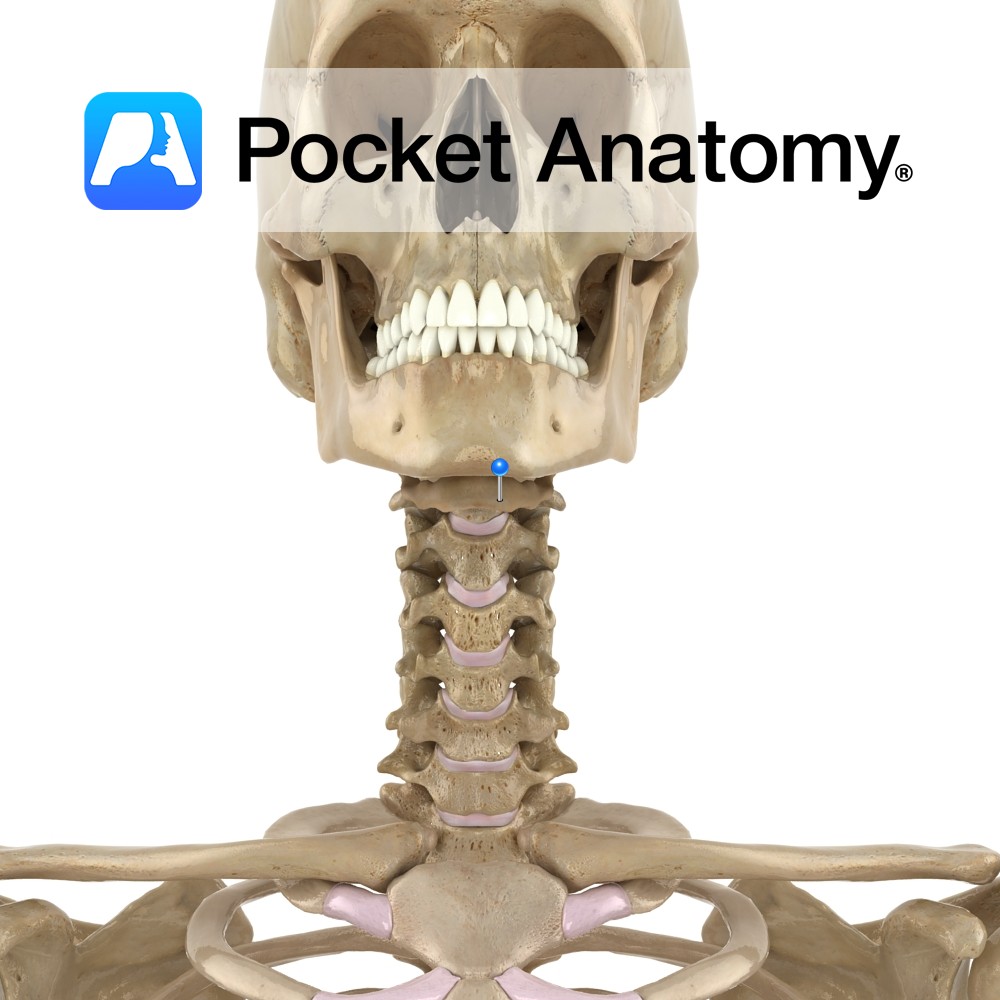
Anatomy Lingual bone, U-shaped, in front of neck, between larynx (behind) and mandible (in front), at level of C3; made up of body (transverse), greater horns/cornus (long, pointing back), lesser cornus/horns (short, pointing up); suspended from tips of styloid processes of mandible by ligaments (this syndesmotic connection being its only bony articulation); anchored by multiple
- Published in Pocket Anatomy Pins
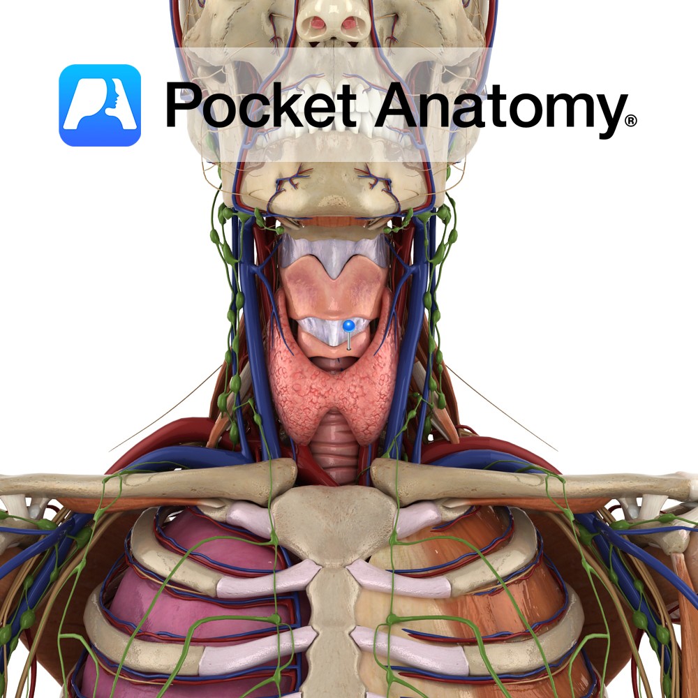
Anatomy The most inferior part of the laryngeal cartilages and completely encircles the trachea. It is shaped like a signet ring with a broad lamina of cricoid cartilage posteriorly and a much slimmer arch of cartilage anteriorly completing the circle. The lamina has two depressions separated by a vertical ridge. There are two articular facets
- Published in Pocket Anatomy Pins
Pocket Anatomy has won the “Boost” StartUp competition at The Next Web (TNW) Conference in Amsterdam. The event is one of Europe’s top tech gatherings and was attended by over 2,500 influential web, technology and business leaders from all over the world. Nearly a hundred StartUps were selected for Boost, out of which 10 (including
- Published in Awards
To celebrate the launch of the new iOS7 operating system update on iPad and iPhone, Pocket Anatomy announces its beautiful and minimalist new interface! <H2>New and Improved</H2> Harnessing the already highly user-friendly 3D interface and features of previous versions, the newest refresh of Pocket Anatomy is a vital addition to anyone’s library of medical Apps.
- Published in App Updates
May 30th sees the launch of one of the most significant medical anatomy apps available on the Apple Platform. The 4th Generation update to the multi award-winning Pocket Anatomy now includes full female and male anatomy. This complements the recent additions of circulatory & lymphatic systems, as well as the inclusion of both cranial &
- Published in App Updates
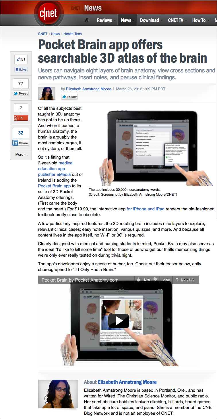
Full text version: Users can navigate eight layers of brain anatomy, view cross sections and nerve pathways, insert notes, and peruse clinical findings. Elizabeth Armstrong Moore by Elizabeth Armstrong Moore March 26, 2012 1:09 PM PDT Of all the subjects best taught in 3D, anatomy has got to be up there. And when it comes
- Published in In the News

Benefits: With Pocket Brain, we’ve made an exciting App that transforms teaching, learning and communication of the human brain. Pocket Brain assists the medical, nursing or health care professionals communicate with their patients, as well as learn and share clinically relevant neuroanatomy with colleagues. For neuroanatomy students, Pocket Brain will help you prepare for and
- Published in App Updates