Motion
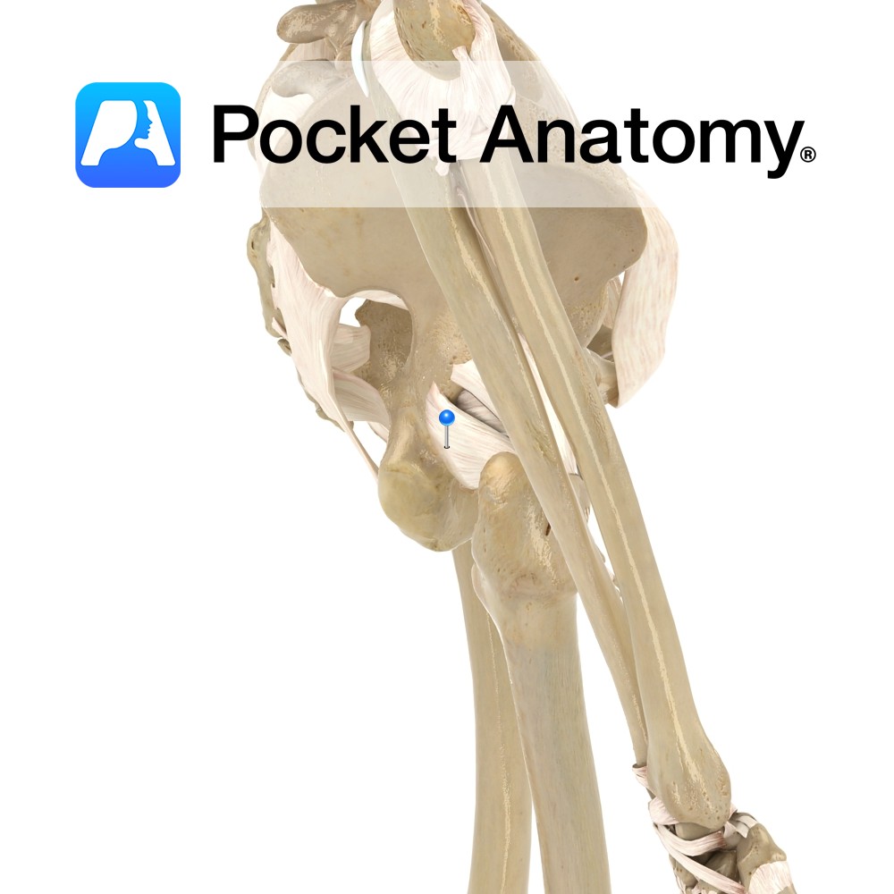
The hip joint is a multiaxial synovial ball and socket joint. It is formed by the articulation between the head of the femur and the acetabulum of the pelvic bone. It can be flexed, extended, abducted, adducted, medially rotated, laterally rotated and circumducted. Circumduction is the complex circular movement of the joint combining flexion, extension, abduction and adduction. The hip joint is designed to take the body’s weight both in standing and dynamic postures.
For this reason the hip has a very high degree of stability, albeit at the expense of some degree of movement. In contrast the shoulder has a higher degree of mobility, but less stability.
Stability
The fibrous capsuleof the hip joint contributes to the joints stability.
The ligaments are important in stabilizing the hip joint and can be divided into extra-capsular and intra-capsular ligaments. The extra-capsular ligaments include:
-Transverse acetabular ligament
–Iliofemoral ligament (anteriorly)
–Ischiofemoral ligament (posteriorly)
–Pubofemoral ligament (anteroinferiorly)
The single intra-capsular ligament includes:
-Ligament of the head of the femor (ligamentum teres femoris) which may contain an arterial supply to the femoral head.
The main muscles involved in stabilizing the hip joint are:
–Psoas major
–Iliacus
–Pectineus
–Rectus femoris
–Gluteus minimus
–Obturator externus
–Obturator internus
–Superior gemellus
–Inferior gemellus
–Piriformis
–Quadratus femoris
The cup shaped acetabulum of the pelvic bone surrounds the head of the femur almost completely. This arrangement plays an important role in joint stability. The acetabular labrum is a ring of fibrocartilage surrounding the rim of the acetabulum. It effectively increases the surface area of the joint and helps prevent the femoral head slipping out of place.
Muscles
Flexion:
Psoas major
Iliacus
Rectus femoris
Sartorius
Pectineus
Tensor faciae latae
Adductor longus (assists)
Extension:
Gluteus maximus
Semitendinosus
Semimembranosus
Biceps femoris (long head)
Abduction:
Pectineus (assists)
Gracilis (assists)
Adductor longus
Adductor brevis
Adductor magnus
Medial Rotation:
Tensor fasciae latae
Semitendinosus
Semimembranosus
Gluteus medius
Gluteus minimus
Adductor longus
Adductor brevis
Adductor magnus
Lateral Rotation:
Psoas major
Iliacus
Sartorius (assists)
Gluteus maximus (assists)
Biceps femoris (long head)
Obturator internus (when femur extended)
Piriformis (when femur extended)
Gemelli (when femur extended)
Obturator externus
Quadratis femoris.
Clinical
Hip replacement surgery is one of the most successful orthopaedic procedures performed today. The aim of the surgery is relief of pain and/or improvement of hip function. Severe arthritis pain, arthritic joint damage and fractures of the femoral neck are some common indications for hip replacement.
Partial hip replacements involve replacing the ‘ball’ (the head of the femur) with a metal prosthesis, is used as a treatment for serious femoral neck fractures. Total hip replacement involves also replacing the ‘socket’ (the acetabulum of the pelvis) and is more typically carried out for arthritic patients.
Interested in taking our award-winning Pocket Anatomy app for a test drive?


.jpg)


